
Pulmonary Function Tests in Children: A Comprehensive Guide to Interpretation and Common Pediatric Diseases
Pulmonary function tests (PFTs) are an important part of evaluating respiratory disease in children. They help clinicians identify airflow obstruction, abnormal lung volumes, air trapping, impaired gas transfer, ventilation inhomogeneity, and exercise limitation.
However, pediatric PFT interpretation is not simply a matter of looking at whether a value is “above or below 80% predicted.” Children are continuously growing, lung volumes change with age and body size, and the ability to perform a technically acceptable test depends strongly on developmental stage.
For postgraduate pediatric examinations and clinical practice, the most useful approach is to understand what each PFT measures, which pattern it produces, and which diseases commonly produce that pattern.
Modern interpretation relies on appropriate reference equations and the lower limit of normal (LLN) rather than applying adult fixed cutoffs to children. The 2022 ERS/ATS interpretive standard also emphasizes interpreting results in the context of test quality and clinical information.
What Are Pulmonary Function Tests?
Pulmonary function tests are a group of investigations used to assess different aspects of respiratory physiology.
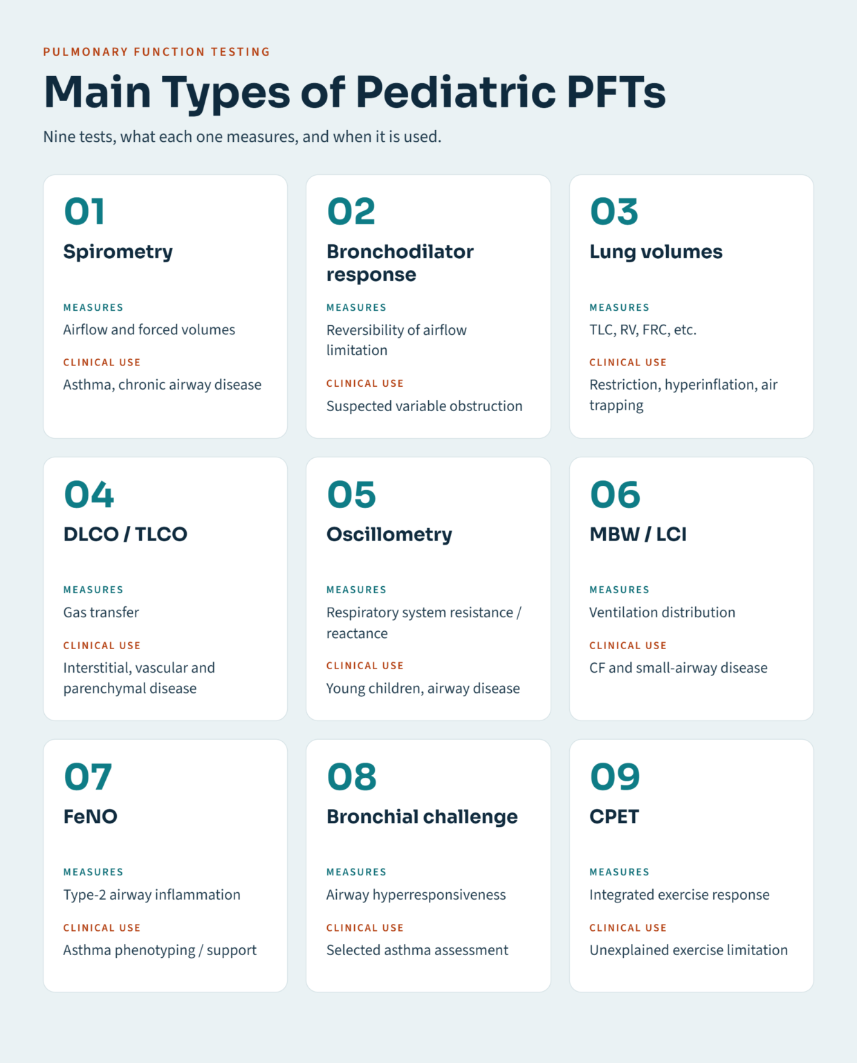
Depending on the child’s age, clinical condition, and ability to cooperate, testing may include:
- Spirometry
- Bronchodilator responsiveness testing
- Lung-volume measurement
- Diffusing capacity for carbon monoxide (DLCO/TLCO)
- Respiratory oscillometry
- Multiple-breath washout (MBW)
- Bronchial challenge testing
- Fractional exhaled nitric oxide (FeNO)
- Exercise testing and cardiopulmonary exercise testing (CPET)
- Specialized infant pulmonary function testing
No single test answers every clinical question.
For example:
Spirometry → airflow
Lung volumes → restriction and hyperinflation
DLCO → gas transfer
Oscillometry → respiratory system mechanics during tidal breathing
MBW/LCI → ventilation inhomogeneity
FeNO → type-2/eosinophilic airway inflammation
CPET → exercise limitation
Difference of Pulmonary Function Tests in Children
Children are not simply small adults.
Lung function changes substantially with:
- Age
- Height
- Growth
- Pubertal development
- Sex
- Body composition
- Reference population
Therefore, pediatric results should be interpreted using an appropriate reference population and laboratory standards.
The correct pediatric approach
For children, pulmonary function results are best interpreted using:
Age- and size-appropriate reference equations
LLN and z-scores
Quality-assured spirometry
Measured lung volumes when restriction is suspected
RV and RV/TLC when air trapping is being assessed
A practical framework is:
| Finding | Pediatric interpretation |
|---|---|
| FEV₁/FVC below LLN | Suggests airflow obstruction |
| FVC below LLN | Indicates reduced forced vital capacity and requires further interpretation |
| TLC below LLN | Confirms a restrictive ventilatory defect |
| RV or RV/TLC increased | Supports air trapping |
| FEV₁/FVC below LLN + TLC below LLN | Mixed obstructive and restrictive physiology |
| DLCO below expected range | Suggests impaired gas transfer, after considering technical and hemoglobin-related factors |
The key principle is that pediatric PFTs should be interpreted against appropriate pediatric reference standards rather than a single fixed adult cutoff.
1.Spirometry
Spirometry is the most commonly used pulmonary function test.
It measures how much air a child can move and how quickly that air can be exhaled.
The major measurements include:
- FVC – forced vital capacity
- FEV₁ – forced expiratory volume in the first second
- FEV₁/FVC – ratio of forced expiratory volume in one second to forced vital capacity
- PEF – peak expiratory flow
- Other forced expiratory flows
FEV₁
FEV₁ is the volume exhaled during the first second of a forced expiration.
It is particularly useful for identifying and quantifying airflow limitation.
A reduced FEV₁ may occur in:
- Obstructive disease
- Restrictive disease
- Mixed disease
- Poor-quality or submaximal effort
Therefore, FEV₁ should never be interpreted in isolation.
FVC
FVC is the total volume forcibly exhaled after a full inspiration.
A reduced FVC can occur with:
- True restriction
- Air trapping
- Incomplete expiration
- Poor effort
- Early termination
- Severe airflow obstruction
Therefore, a reduced FVC should prompt consideration of the complete physiological pattern.
When restriction is suspected, TLC is the measurement used to establish whether restriction is actually present.
FEV₁/FVC
The FEV₁/FVC ratio is particularly useful for identifying airflow obstruction.
A value below the age-appropriate LLN suggests an obstructive ventilatory defect.
The LLN is generally related to the lower fifth percentile of the reference population, corresponding approximately to a z-score of −1.645 when the reference model is appropriately constructed.
Thus, in children:
Use the age-appropriate LLN to identify airflow obstruction.
The Flow-Volume Loop
The flow-volume loop provides visual information about expiratory and inspiratory airflow.
Obstructive pattern
Typically:
- Reduced expiratory flow
- Concave or “scooped” expiratory limb
- Reduced FEV₁/FVC
Common examples include:
- Asthma
- Cystic fibrosis
- Bronchiectasis
- Bronchiolitis obliterans
Restrictive pattern
A restrictive ventilatory defect generally produces:
- Reduced lung volumes
- Relatively preserved or increased FEV₁/FVC
- A smaller but proportionally shaped loop
However, spirometry alone cannot reliably confirm restriction.
Upper-airway obstruction
Characteristic flattening may occur depending on whether obstruction is:
- Variable extrathoracic
- Variable intrathoracic
- Fixed
This is an important additional use of the flow-volume loop.
Pediatric Spirometry: Always Check Test Quality First
Before interpreting numbers, ask:
1. Was the maneuver acceptable?
Consider:
- Good start of test
- Adequate inspiration
- Rapid maximal expiration
- No cough during critical portions
- No glottic closure
- No leak
- No early termination
2. Are the maneuvers repeatable?
The best results should be supported by appropriately reproducible maneuvers according to current technical standards.
3. Is the child able to perform the test?
Younger children may require extensive coaching.
Importantly, preschool children are not automatically unable to perform spirometry. Many can produce useful measurements with appropriate coaching, although feasibility and quality vary considerably.
Spirometry by Age
Infants
Routine conventional spirometry is generally not feasible in an awake infant.
Specialized infant pulmonary function laboratories can perform techniques such as:
- Tidal breathing analysis
- Infant forced expiratory maneuvers
- Respiratory mechanics
- Infant plethysmography
These techniques require specialized equipment and expertise.
Preschool Children
Some preschool children can perform acceptable spirometry, particularly with experienced coaching.
When forced maneuvers are difficult or unreliable, other techniques may be useful, including:
- Oscillometry
- Multiple-breath washout
- Other specialized preschool lung-function techniques
Therefore, the appropriate statement is:
Spirometry can be performed successfully in many preschool children, while oscillometry and other tidal-breathing techniques provide useful alternatives when forced maneuvers are difficult.
School-Aged Children and Adolescents
Standard spirometry is generally feasible in most cooperative school-aged children and adolescents.
At this age, interpretation should emphasize:
- Test quality
- Reference equations
- FEV₁/FVC
- FEV₁
- FVC
- Bronchodilator response when indicated
- Flow-volume loop morphology
A Practical Approach to Spirometry Interpretation
A useful postgraduate approach is:
Step 1: Check test quality
Confirm that the results are technically reliable.
Step 2: Look at FEV₁/FVC
- Below LLN → suggests obstruction
- Within the expected range → obstruction is not demonstrated by the ratio
Step 3: Assess FVC
- Within expected range → obstruction may be isolated
- Reduced → consider the complete pattern, including restriction, air trapping and test quality
Step 4: If restriction is suspected, measure TLC
Reduced TLC → confirms a restrictive ventilatory defect.
Step 5: Examine bronchodilator response when clinically indicated
A meaningful response may support variable airflow limitation, but absence of a response on one occasion does not exclude asthma.
Step 6: Integrate the findings
PFTs describe physiological abnormalities; the final diagnosis requires clinical correlation.
Obstructive Ventilatory Defect
The typical pattern is:
- FEV₁/FVC below LLN
- FEV₁ may be reduced
- FVC may be normal or reduced
- Expiratory limb may show concavity
Common pediatric causes include:
- Asthma
- Cystic fibrosis
- Bronchiectasis
- Bronchiolitis obliterans
- Primary ciliary dyskinesia
- Chronic airway disease after severe early-life lung injury
Important exam point
A reduced FEV₁/FVC identifies airflow obstruction, not a specific disease.
Clinical history is required to determine the cause.
Restrictive Ventilatory Defect
A restrictive defect means that the total amount of air contained in the lungs is reduced.
The key physiological finding is:
Reduced TLC
Spirometry may suggest restriction when:
- FVC is reduced
- FEV₁ is reduced proportionally
- FEV₁/FVC is preserved or increased
But:
Confirmation comes from demonstrating reduced TLC.
Potential causes include:
- Interstitial lung disease
- Chest-wall disorders
- Severe neuromuscular weakness
- Severe obesity
- Pleural disease
- Certain developmental lung disorders
Mixed Ventilatory Defect
A mixed defect combines:
- Obstruction → low FEV₁/FVC
- Restriction → low TLC
Therefore, a genuinely mixed pattern requires evidence of both abnormalities.
A low FEV₁/FVC combined with low FVC on spirometry should be further evaluated with lung-volume measurement to determine whether true restriction is present.
This distinction is particularly important in examination questions.
Air Trapping and Hyperinflation
Lung-volume testing can reveal abnormalities that spirometry may not fully demonstrate.
Important measurements include:
- RV – residual volume
- FRC – functional residual capacity
- TLC – total lung capacity
Air trapping
Usually supported by increased:
RV
or
RV/TLC
Hyperinflation
May be associated with an increased:
TLC
and/or increased FRC, depending on the underlying physiology.
Air trapping is particularly relevant in:
- Asthma
- Bronchiolitis obliterans
- Cystic fibrosis
- Bronchiectasis
- Small-airway disease
Bronchodilator Responsiveness
Bronchodilator testing assesses whether airflow changes after administration of a short-acting bronchodilator.
The modern ERS/ATS interpretive approach uses change relative to the predicted value rather than relying on the older adult criterion of “>12% and >200 mL.”
The 2022 ERS/ATS approach defines a significant bronchodilator response as an increase of more than 10% of the predicted value in FEV₁ or FVC.
The result must still be interpreted in clinical context.
Important point
A negative bronchodilator response does not by itself exclude asthma.
Similarly, a positive response does not independently establish asthma.
2. Lung Volumes
Spirometry cannot measure all lung volumes.
Important lung-volume measurements include:
Total lung capacity (TLC)
The total amount of air in the lungs after maximal inspiration.
Reduced TLC → confirms restriction.
Residual volume (RV)
The air remaining in the lungs after maximal expiration.
Increased RV → supports air trapping.
Functional residual capacity (FRC)
The volume remaining in the lungs at the end of normal expiration.
It may increase with hyperinflation and decrease in conditions that reduce resting lung volume.
RV/TLC
An increased RV/TLC ratio supports air trapping.
How Lung Volumes Help Solve Difficult Spirometry
Consider a child with:
- Low FEV₁
- Low FVC
- Low FEV₁/FVC
You might initially think:
Mixed obstructive and restrictive disease.
But lung-volume testing could show:
- TLC normal
- RV increased
This suggests obstruction with air trapping rather than true restriction.
Therefore:
TLC is the key measurement for confirming restriction.
3. Diffusing Capacity: DLCO/TLCO
DLCO assesses the transfer of gas from the alveoli into pulmonary capillary blood.
It depends on several factors, including:
- Alveolar-capillary surface area
- Membrane characteristics
- Pulmonary capillary blood volume
- Hemoglobin concentration
A reduced DLCO may be seen in selected:
- Interstitial lung diseases
- Pulmonary vascular diseases
- Emphysematous disorders
- Conditions associated with anemia
DLCO should be interpreted with attention to hemoglobin and technical factors.
Pediatric exam pearl
If a child has:
Restriction + reduced DLCO
think particularly about intrinsic/parenchymal lung disease.
If there is:
Restriction + relatively preserved DLCO
consider extraparenchymal causes such as:
- Chest-wall restriction
- Neuromuscular weakness
- Some obesity-related patterns
This is a useful pattern-recognition approach, but individual diseases may vary.
4. Respiratory Oscillometry
Oscillometry measures respiratory system mechanics during normal tidal breathing.
It is especially useful when a child cannot reliably perform forced expiratory maneuvers.
Parameters can include:
- Resistance
- Reactance
- Frequency dependence
- Area under the reactance curve
Oscillometry can be particularly useful in:
- Preschool children
- Asthma
- Cystic fibrosis
- Other chronic airway diseases
Why is it useful in young children?
The child generally breathes normally through the device rather than performing repeated maximal forced expirations.
Therefore, it can complement—not necessarily replace—spirometry.
5. Multiple-Breath Washout and Lung Clearance Index
Multiple-breath washout (MBW) assesses how evenly ventilation is distributed throughout the lungs.
One important outcome is:
Lung clearance index (LCI)
An elevated LCI indicates increased ventilation inhomogeneity.
MBW can detect abnormalities that may occur before conventional spirometry becomes clearly abnormal.
It has particular clinical and research importance in:
- Cystic fibrosis
- Small-airway disease
- Preschool children
The ATS preschool MBW technical statement specifically addresses the methodological challenges of performing MBW in young children.
Important distinction
MBW does not directly measure airway obstruction in the same way as FEV₁/FVC.
Instead, it assesses ventilation distribution and inhomogeneity.
6. Fractional Exhaled Nitric Oxide (FeNO)
FeNO is a marker associated with type-2/eosinophilic airway inflammation.
It can support the assessment of asthma, particularly when eosinophilic inflammation is suspected.
However:
FeNO is not a stand-alone diagnostic test for asthma.
FeNO may be influenced by:
- Atopy
- Allergic rhinitis
- Inhaled corticosteroid treatment
- Age
- Smoking exposure
- Other inflammatory conditions
Therefore, FeNO should be interpreted alongside symptoms, spirometry and the overall clinical picture.
7. Bronchial Challenge Testing
Bronchial challenge tests evaluate airway hyperresponsiveness.
Depending on the laboratory and clinical question, challenges may involve:
- Exercise
- Methacholine
- Other standardized stimuli
They can be useful when asthma is suspected but baseline spirometry is nondiagnostic.
However, a challenge test should be selected and interpreted according to the clinical context and laboratory standards.
8. Exercise Testing and CPET
Some children have significant exercise intolerance despite relatively normal resting spirometry.
Cardiopulmonary exercise testing (CPET) evaluates the integrated response of:
- Respiratory system
- Cardiovascular system
- Muscles
- Gas exchange
- Exercise capacity
It can help investigate:
- Unexplained exertional dyspnea
- Exercise limitation in chronic lung disease
- Congenital heart disease
- Obesity
- Dysfunctional breathing
- Deconditioning
Pulmonary Function Tests in Children ( Disease-Wise Interpretation)
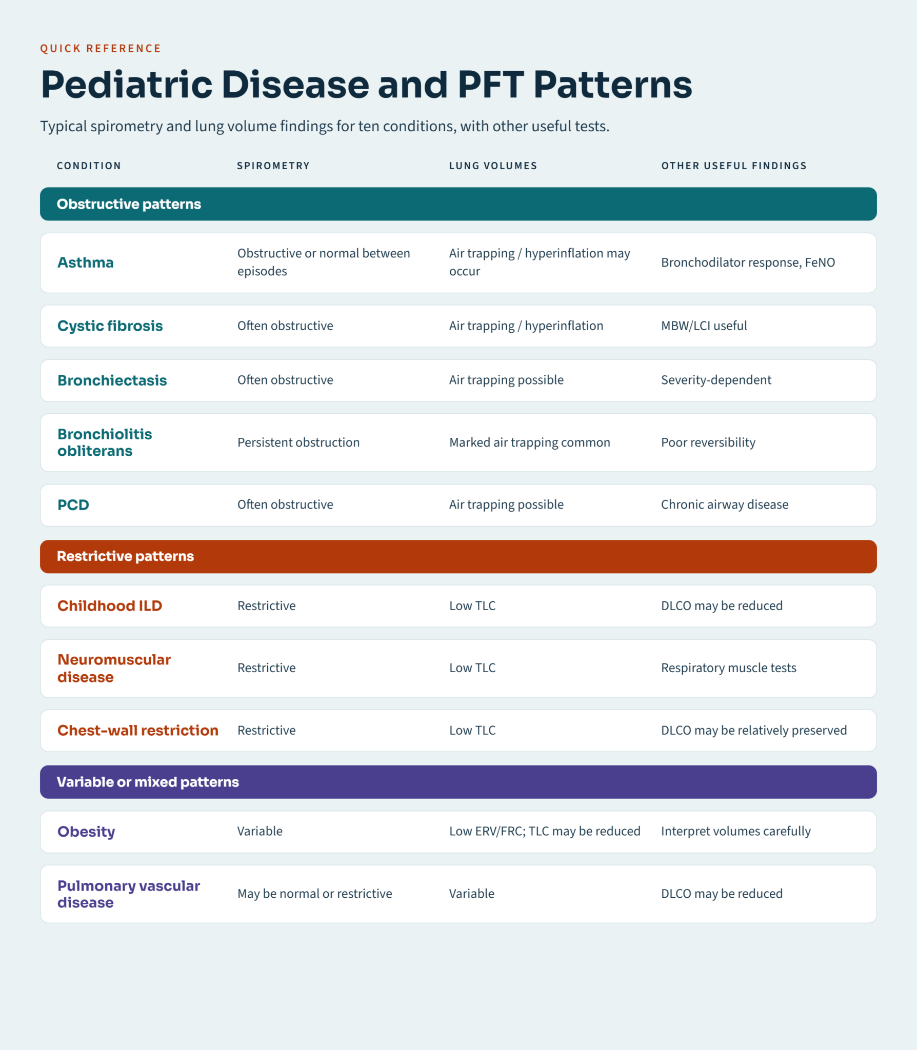
The following patterns are useful for postgraduate examinations, but they should be regarded as typical patterns rather than absolute diagnostic rules.
Asthma
Possible findings:
- Normal spirometry between episodes
- Reduced FEV₁/FVC during airflow obstruction
- Reduced FEV₁
- Bronchodilator responsiveness in some patients
- Air trapping or hyperinflation in more persistent disease
- Increased FeNO in some patients with type-2 inflammation
Key point
Normal spirometry does not automatically exclude asthma, particularly when symptoms are intermittent.
Cystic Fibrosis
Possible findings include:
- Obstructive physiology
- Reduced FEV₁
- Air trapping
- Hyperinflation
- Progressive decline in lung function
- Ventilation inhomogeneity on MBW
LCI/MBW may provide additional information, particularly in younger children.
Exam pearl
In CF:
A normal FEV₁ does not necessarily mean completely normal peripheral airway function.
This is one reason MBW and LCI have been studied extensively in pediatric CF.
Bronchiectasis
Possible findings:
- Obstructive physiology
- Reduced FEV₁
- Air trapping
- Sometimes mixed abnormalities
The pattern depends on disease severity, extent and underlying cause.
Possible underlying conditions include:
- Cystic fibrosis
- Primary ciliary dyskinesia
- Immunodeficiency
- Previous severe infection
- Other chronic suppurative lung diseases
Bronchiolitis Obliterans
A typical pattern is:
- Persistent airflow obstruction
- Reduced FEV₁/FVC
- Reduced FEV₁
- Significant air trapping
- Possible hyperinflation
The obstruction is generally poorly reversible.
This pattern is particularly important after severe lower respiratory tract injury or hematopoietic stem-cell transplantation.
Primary Ciliary Dyskinesia
PFT abnormalities may include:
- Airflow obstruction
- Reduced FEV₁
- Air trapping
- Progressive decline in lung function
However, PFTs are not diagnostic of PCD.
Diagnosis requires integration of clinical phenotype with specialized diagnostic testing.
Post-Prematurity Respiratory Disease
Children and adolescents born very preterm may demonstrate heterogeneous lung-function abnormalities.
Possible findings include:
- Airflow obstruction
- Reduced lung volumes in some patients
- Air trapping
- Abnormal respiratory mechanics
- Exercise limitation
The phenotype varies according to gestational age, neonatal lung disease, oxygen exposure, mechanical ventilation and subsequent development.
Childhood Interstitial Lung Disease
PFTs may show:
- Reduced FVC
- Reduced TLC
- Restrictive physiology
- Reduced DLCO in some disorders
A restrictive pattern with impaired gas transfer can support the presence of significant parenchymal lung disease.
However, PFTs should be interpreted with imaging, clinical findings and other investigations.
Neuromuscular Disease
Respiratory muscle weakness commonly produces:
- Reduced FVC
- Restrictive physiology
- Reduced TLC
- Difficulty generating adequate expiratory force
Additional assessment may include:
- Maximal inspiratory pressure
- Maximal expiratory pressure
- Peak cough flow
- Supine versus upright FVC
- Sleep-related respiratory assessment when indicated
An increased RV may occur when expiratory muscle weakness impairs effective emptying.
Obesity
Obesity can alter respiratory mechanics.
Common findings include:
- Reduced ERV
- Reduced FRC
- Sometimes reduced TLC
- Increased airway resistance in some patients
Importantly:
Reduced FRC or ERV does not by itself prove restrictive lung disease.
True restriction requires a reduced TLC.
A 7-Step Pediatric PFT Interpretation Algorithm
When faced with a PFT question in an examination, use the following sequence:
Step 1 — Confirm test quality
Make sure the study is technically reliable.
Step 2 — Assess FEV₁/FVC
Below LLN → airflow obstruction is suggested.
Step 3 — Assess FVC
Determine whether FVC is within the expected range or reduced, then consider the complete physiological pattern.
Step 4 — Check TLC when restriction is suspected
Low TLC → restriction confirmed.
Step 5 — Check RV and RV/TLC
Increased RV or RV/TLC → supports air trapping.
Step 6 — Consider bronchodilator response
A significant response may support variable airflow limitation.
Step 7 — Integrate with the clinical picture
Ask:
- What is the child’s age?
- What are the symptoms?
- Is disease episodic or persistent?
- Is there atopy?
- Is there chronic infection?
- Was the child born prematurely?
- Is there neuromuscular weakness?
- Are imaging findings available?
High-Yield Pediatric PFT Examination Points
Point 1
Use age-appropriate reference equations and LLN when interpreting pediatric spirometry.
Point 2
Use TLC to establish restriction.
Point 3
Mixed physiology = obstruction plus confirmed restriction.
That means:
Low FEV₁/FVC + low TLC
Point 4
Use RV and RV/TLC to recognize air trapping.
Point 5
Normal spirometry does not exclude asthma.
Asthma is characterized by variable respiratory symptoms and variable airflow limitation.
Point 6
Interpret FEF25–75 cautiously and never use it alone to diagnose small-airway disease.
Point 7
Preschool children can sometimes perform useful spirometry.
Point 8
Oscillometry is particularly useful when forced maneuvers are difficult.
Point 9
MBW/LCI assesses ventilation inhomogeneity.
Point 10
PFTs describe physiology, not the diagnosis by themselves.
Always integrate PFT findings with clinical history, examination and other investigations.
The Simplest Way to Remember Pediatric PFTs
Think of pulmonary function testing as answering five questions:
1. Is airflow obstructed?
Look at:
FEV₁/FVC
2. Are the lungs too small?
Look at:
TLC
3. Is air trapped?
Look at:
RV and RV/TLC
4. Is gas transfer impaired?
Look at:
DLCO/TLCO
5. Is ventilation uneven?
Think about:
MBW and LCI
This framework makes complicated PFT reports much easier to understand.
Final Clinical Summary
Pediatric PFT interpretation should begin with test quality and appropriate reference equations, followed by assessment of airflow, lung volumes and gas transfer where available.
The core pattern recognition is:
Low FEV₁/FVC → obstruction
Low TLC → restriction
Low FEV₁/FVC + low TLC → mixed physiology
High RV/RV/TLC → air trapping
Low DLCO → impaired gas transfer
High LCI → ventilation inhomogeneity
The most important principle is that these patterns are physiological descriptions, not diagnoses. Asthma, cystic fibrosis, bronchiectasis, bronchiolitis obliterans, primary ciliary dyskinesia, childhood interstitial lung disease, neuromuscular disorders and obesity can produce overlapping patterns.
For postgraduate pediatric examinations, the safest strategy is therefore to interpret the PFT systematically rather than trying to memorize one fixed pattern for every disease.
References
- Stanojevic S, Kaminsky DA, Miller MR, et al. ERS/ATS technical standard on interpretive strategies for routine lung function tests. Eur Respir J. 2022;60(1):2101499.
- Graham BL, Steenbruggen I, Miller MR, et al. Standardization of Spirometry 2019 Update. An Official American Thoracic Society and European Respiratory Society Technical Statement. Am J Respir Crit Care Med. 2019;200(8).
- Beydon N, Davis SD, Lombardi E, et al. An official American Thoracic Society/European Respiratory Society statement: pulmonary function testing in preschool children. Am J Respir Crit Care Med. 2007;175:1304-1345.
- Robinson PD, Latzin P, Ramsey KA, et al. Preschool Multiple-Breath Washout Testing. An Official American Thoracic Society Technical Statement. Am J Respir Crit Care Med. 2018;197(5).
- Culver BH, Graham BL, Coates AL, et al. Recommendations for a Standardized Pulmonary Function Report. An Official American Thoracic Society Technical Statement. Am J Respir Crit Care Med. 2017;196(11):1463-1472.
- Quanjer PH, Stanojevic S, Cole TJ, et al. Multi-ethnic reference values for spirometry for the 3-95-yr age range: the Global Lung Function 2012 equations. Eur Respir J. 2012;40(6):1324-1343.
