Clinical Scenario:
A 12-year-old boy presents with progressive abdominal distension and reduced exercise tolerance for 4 months. There is no history of jaundice or previous liver disease.
On examination, his blood pressure is 90/60 mmHg, pulse rate is 96/min, and the jugular venous pressure is elevated and rises with inspiration. He has marked ascites, hepatomegaly and bilateral ankle edema. Heart sounds are relatively quiet, and an early diastolic pericardial knock is audible.
Chest radiograph shows a normal-sized cardiac silhouette with pericardial calcification.
Which of the following is the most likely diagnosis?
A. Chronic liver disease
B. Dilated cardiomyopathy
C. Constrictive pericarditis
D. Nephrotic syndrome
E. Restrictive cardiomyopathy
Correct Answer & Explanation:
Correct answer: C. Constrictive pericarditis
Explanation
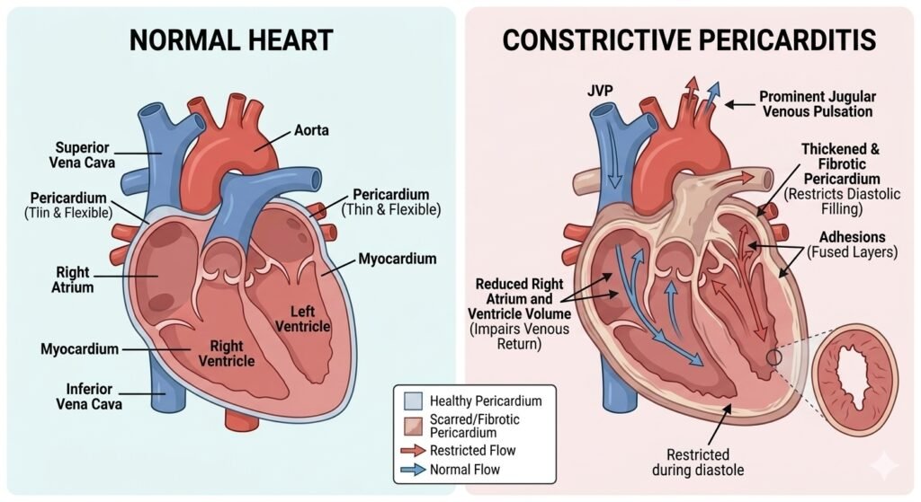
This child has predominantly right-sided heart failure caused by impaired ventricular filling, with marked systemic venous congestion manifested by raised JVP, hepatomegaly, ascites and peripheral edema.
The most important clinical clue is the JVP rising with inspiration (Kussmaul sign). In constrictive pericarditis, the pericardium becomes thickened, fibrotic and non-compliant. During inspiration, systemic venous return increases, but the rigid pericardium prevents the right ventricle from expanding normally. Consequently, venous pressure rises rather than falling, producing the characteristic Kussmaul sign.
Another important finding is the early diastolic pericardial knock. This is a high-pitched early diastolic sound produced by the abrupt cessation of ventricular filling when the expanding ventricles rapidly encounter the stiff, non-compliant pericardium. It can be an important clue distinguishing constrictive pericarditis from other causes of right-sided heart failure.
The low blood pressure (90/60 mmHg) may reflect reduced ventricular filling and consequently reduced stroke volume and cardiac output. However, hypotension is a supportive finding rather than a diagnostic feature of constrictive pericarditis.
The normal-sized cardiac silhouette despite substantial systemic venous congestion is another useful clue. Unlike many patients with dilated cardiomyopathy, the heart is not necessarily enlarged because the principal abnormality is impaired filling caused by the constricting pericardium rather than primary ventricular dilation.
Pericardial calcification, if present, strongly supports chronic constrictive pericardial disease. However, its absence does not exclude constrictive pericarditis. Echocardiography, CT or cardiac MRI may be required when the clinical diagnosis is uncertain.
In a child living in or originating from a tuberculosis-endemic setting, previous tuberculous pericarditis should be considered an important possible cause. Other causes include previous cardiac surgery, bacterial or viral pericarditis, radiation and other inflammatory conditions.
Why not restrictive cardiomyopathy?
Restrictive cardiomyopathy can closely mimic constrictive pericarditis because both conditions cause impaired diastolic filling, elevated systemic venous pressure, hepatomegaly, ascites and peripheral edema, often with relatively preserved systolic function.
However, the pericardial knock and evidence of pericardial thickening or calcification favor constrictive pericarditis. In restrictive cardiomyopathy, the primary abnormality is within the myocardium, whereas in constrictive pericarditis the abnormality is predominantly in the pericardium.
Why the other diagnoses are less likely
Dilated cardiomyopathy: Usually produces ventricular dilation and predominantly systolic dysfunction. A normal-sized heart with marked systemic venous congestion and a pericardial knock is less consistent with this diagnosis.
Right ventricular failure due to pulmonary hypertension: Can cause raised JVP, hepatomegaly and edema, but the pericardial knock and pericardial calcification point toward a pericardial cause rather than primary pulmonary vascular disease.
Cardiac tamponade: Also causes impaired cardiac filling and systemic venous congestion, but tamponade is generally an acute or subacute process and is characterized by pericardial fluid under pressure rather than a chronically rigid pericardium. A pericardial knock is not typical of tamponade.
Severe tricuspid regurgitation: Produces prominent systemic venous congestion and may cause a large V wave in the JVP, hepatomegaly and edema. However, the characteristic early diastolic pericardial knock and pericardial calcification favor constrictive pericarditis.
Clinical Pearl
Think of constrictive pericarditis when severe systemic venous congestion occurs without cardiomegaly, particularly when there is a Kussmaul sign and an early diastolic pericardial knock.
A useful exam distinction is:
Constrictive pericarditis → abnormal pericardium
Restrictive cardiomyopathy → abnormal myocardium
Both can produce predominantly right-sided heart failure with preserved ejection fraction, so the pericardial findings and imaging are particularly useful in distinguishing them.
High-Yield Exam Point
Kussmaul sign + pericardial knock + systemic venous congestion + normal-sized heart ± pericardial calcification = strongly consider constrictive pericarditis.
“Related Practice: Test your skills with our [MCQ on Acute Rheumatic Fever] to master pediatric cardiology.”
Reference:
Schulz-Menger J, Collini V, Gröschel J, et al. 2025 ESC Guidelines for the management of myocarditis and pericarditis. European Heart Journal. 2025;46(40):3952–4041. [Read the ESC guideline]