Congenital heart diseases | Neonatal Presentation

Neonates who present with congenital heart disease

Its a common and high yield exam question . Usually a neonate present in a very critical situation and all the Clinical scenarios are overlapping. Here we will learn how to differentiate.ย 

Hypoplastic Left Heart Syndrome (HLHS) โ€” Neonatal Presentation

  • Cyanosis soon after birth, often worsening as the ductus arteriosus closes.
  • Poor feeding, lethargy, weak cry.
  • Tachypnea and respiratory distress.
  • Poor pulses, especially peripheral pulses.
  • Shock / poor perfusion: cold extremities, prolonged capillary refill.
  • Metabolic acidosis due to poor systemic blood flow.
  • May develop heart failure with hepatomegaly and pulmonary edema.

Key exam clue:
๐Ÿ‘‰ A neonate who becomes severely ill, pale/cyanotic and shocked when the ductus closes โ†’ think HLHS.

Important: In HLHS, ductal patency is life-saving โ†’ start prostaglandin Eโ‚ (PGEโ‚) urgently.

ย 

Tricuspid Atresia โ€” Typical Neonatal Presentation

  • Cyanosis โ€” often present from birth or develops early.
  • Tachypnea / respiratory distress.
  • Poor feeding, lethargy and signs of low cardiac output.
  • Single loud S2 may be heard.
  • Murmur due to associated VSD or pulmonary outflow obstruction.
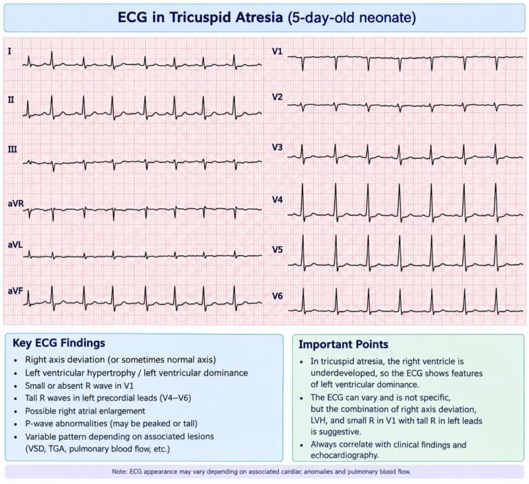
  • ECG: classically left axis deviation and LVH.
  • Severity depends on pulmonary blood flow:
    • Reduced pulmonary flow โ†’ severe cyanosis
    • Increased pulmonary flow โ†’ heart failure

Key exam clue:
๐Ÿ‘‰ Neonate with cyanosis + single S2 + left axis deviation/LVH โ†’ think tricuspid atresia.

TGA with Intact Ventricular Septum โ€” Neonatal Presentation

  • Severe cyanosis immediately after birth โ€” often the main feature.
  • Tachypnea but usually no marked respiratory distress initially.
  • Poor feeding, lethargy and irritability.
  • Minimal or no murmur.
  • Single loud S2 may be heard.
  • Severe hypoxemia that responds poorly to oxygen.
  • ECG: may be normal or show right ventricular dominance.
  • Rapid deterioration if PDA/atrial mixing is inadequate.

Key exam clue:
๐Ÿ‘‰ A profoundly cyanotic neonate with little/no murmur and poor response to oxygen โ†’ think TGA with intact ventricular septum.

Management clue: Maintain PDA with PGEโ‚ and, if atrial mixing is inadequate, perform urgent balloon atrial septostomy (Rashkind procedure).

Critical Coarctation of Aorta โ€” Neonatal Presentation

  • Often normal at birth, while the PDA is open.
  • Deteriorates when PDA closes โ†’ usually in the first daysโ€“weeks.
  • Poor feeding, lethargy, tachypnea.
  • Heart failure and shock.
  • Weak/absent femoral pulses.
  • Upper limb BP > lower limb BP.
  • Metabolic acidosis due to poor systemic perfusion.
  • May have differential cyanosis โ€” lower body may become cyanotic.

Key exam clue:
๐Ÿ‘‰ Neonate who becomes shocked after PDA closure + weak femoral pulses + upper-limb hypertension โ†’ critical coarctation.

Emergency: Start PGEโ‚ to reopen/maintain the PDA and restore systemic blood flow.

ย 

The shortest exam framework

BLUE baby โ†’

  • TGA
  • Pulmonary atresia
  • Critical pulmonary stenosis
  • Tricuspid atresia
  • Obstructed TAPVR
  • Severe Ebstein anomaly

SHOCK baby โ†’

  • HLHS
  • Critical coarctation
  • Interrupted aortic arch
  • Critical aortic stenosis

BLUE + pulmonary edema/respiratory distress โ†’

  • Obstructed TAPVR

SHOCK + weak femoral pulses โ†’

  • Coarctation / interrupted aortic arch

Profound cyanosis + little/no murmur โ†’

  • TGA with intact septum

One important correction to my earlier answer: TGA is primarily a mixing-dependent lesion, not simply a duct-dependent pulmonary-flow lesion. An adequate atrial communication is particularly important, and PGEโ‚ may help maintain ductal mixing, but restrictive atrial mixing requires urgent balloon atrial septostomy.

๐Ÿ‘‰ Neonate with cyanosis + single S2 + left axis deviation/LVH โ†’ think tricuspid atresia.

๐Ÿ‘‰ A neonate who becomes severely ill, pale/cyanotic and shocked when the ductus closes โ†’ think Hypoplastic left heart syndromeย 

๐Ÿ‘‰ Very blue baby + relatively good pulses/perfusion + little/no murmur. Think of Transposition of great arteries with intact septumย 

๐Ÿ‘‰ Neonate who becomes shocked after PDA closure + weak femoral pulses + upper-limb hypertension โ†’ critical coarctation.

Table to make differentials :

Lesion

Main presentation

Pulses

BP

Cyanosis

Key clue

TGA + intact septum

Severe cyanosis

Usually good

Usually maintained

Marked

Very blue baby, little/no murmur

HLHS

Shock + heart failure

Weak all over

Low

Mildโ€“moderate

Poor perfusion + metabolic acidosis

Critical coarctation

Shock + heart failure

Weak/absent femoral

Arm > leg

Usually mild initially

Pulse/BP difference

Critical pulmonary stenosis/atresia

Severe cyanosis

Usually good

Usually maintained

Marked

Little pulmonary blood flow; murmur may be absent

Pulmonary atresia + IVS

Profound cyanosis

Usually good

Usually maintained

Marked

Severe cyanosis from birth; very little pulmonary flow

TAPVR obstructed

Severe cyanosis + respiratory distress

Usually present

May fall

Marked

Pulmonary edema, severe respiratory distress

Interrupted aortic arch

Shock after PDA closure

Upper pulses > lower

Arm > leg

Variable

Differential pulses + severe acidosis

Scroll to Top