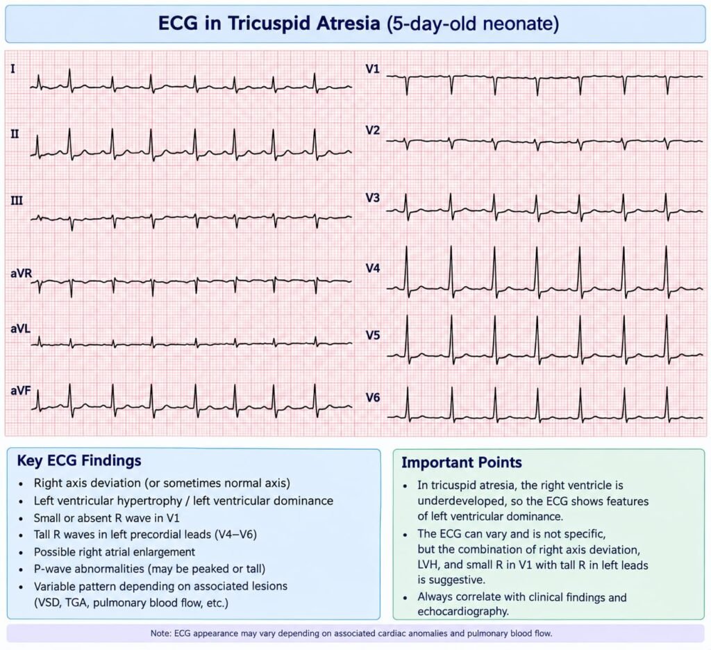
Clinical Scenario:
A 2-day-old neonate develops progressively worsening central cyanosis. He was relatively stable immediately after birth, but the cyanosis has become more marked over the last 24 hours. Oxygen saturation is 70% in room air and shows little improvement with supplemental oxygen. He is tachypnoeic but has no significant chest retractions.
On examination, there is a single loud second heart sound and a harsh pansystolic murmur at the lower left sternal border. Peripheral pulses are palpable and equal in all four limbs. There are no clinical features of pulmonary edema.
ECG shows marked left-axis deviation. Chest radiograph demonstrates reduced pulmonary vascular markings with a normal or mildly increased cardiac silhouette.
Which of the following is the most likely diagnosis?
A. Critical pulmonary stenosis
B. D-transposition of the great arteries
C. Tetralogy of Fallot
D. Tricuspid atresia
E. Total anomalous pulmonary venous connection
Correct Answer & Explanation:
The most likely diagnosis is D. Tricuspid atresia.
Explanation: Tricuspid Atresia
The combination of:
- Progressive cyanosis after birth → suggests a duct-dependent or mixing-dependent congenital heart lesion.
- Single loud S2 → characteristic of tricuspid atresia because there is no normal tricuspid valve contribution to right ventricular flow.
- Harsh pansystolic murmur at the lower left sternal border → usually from an associated VSD.
- Marked left-axis deviation on ECG → a particularly important clue for tricuspid atresia. This is due to the abnormal ventricular activation pattern with a dominant left ventricle.
- Reduced pulmonary vascular markings → consistent with reduced pulmonary blood flow, especially when pulmonary stenosis/atresia or a restrictive VSD limits pulmonary flow.
- No pulmonary edema → argues against lesions with excessive pulmonary blood flow, such as unobstructed TAPVC.
- Equal peripheral pulses → makes a major systemic outflow obstruction less likely.
Why the other options are less likely
A. Critical pulmonary stenosis
Usually produces a right-axis deviation or right ventricular strain pattern rather than marked left-axis deviation.
B. D-transposition of the great arteries
Typically presents with severe cyanosis early after birth, but the ECG does not characteristically show marked left-axis deviation. The murmur may also be absent unless an associated VSD or outflow obstruction is present.
C. Tetralogy of Fallot
Can cause cyanosis and reduced pulmonary vascular markings, but the ECG typically shows right-axis deviation and right ventricular hypertrophy, not marked left-axis deviation.
E. Total anomalous pulmonary venous connection
Obstructed TAPVC can cause profound cyanosis with reduced pulmonary venous drainage, but pulmonary vascular markings are generally increased in unobstructed TAPVC and the clinical picture differs. A single S2 plus marked left-axis deviation strongly favors tricuspid atresia.
High-yield exam clue
Neonate with cyanosis + single S2 + VSD murmur + marked left-axis deviation + reduced pulmonary blood flow = think tricuspid atresia.
Reference:
Minocha PK, Horenstein MS, Phoon C. Tricuspid Atresia. StatPearls [Internet]. StatPearls Publishing.Differential Diagnosis Tip: When evaluating a neonate presenting with cardiogenic shock, it is critical to rule out other left-sided obstructive lesions. Test your diagnostic skills with our practice question on [Coarctation of the Aorta Neonatal Presentation].
ECG