
Ulcerative Colitis — Diagnostic Features
Think of ulcerative colitis when a child has:
- Chronic bloody diarrhea
- Increased stool frequency
- Urgency
- Tenesmus
- Abdominal cramping
- Continuous colonic inflammation
- Disease typically beginning at the rectum
Typical endoscopic findings
- Diffuse erythema
- Loss of normal vascular pattern
- Friability
- Granularity
- Superficial ulceration
- Continuous involvement
Histology
Typical chronic inflammatory changes include:
- Crypt architectural distortion
- Cryptitis
- Crypt abscesses
- Chronic inflammatory infiltrate
- Goblet-cell/mucus depletion
Exam caution: crypt abscesses support active colitis but are not specific for UC.
Pediatric exception
Children can develop atypical UC, including rectal sparing, patchy inflammation, upper-GI involvement and backwash ileitis. Therefore, an atypical distribution does not automatically mean Crohn disease.
You can also practice MCQ on Ulcerative Colitis.
Crohn Disease — Diagnostic Features
Think of Crohn disease when a child has:
- Chronic abdominal pain
- Weight loss
- Growth failure
- Delayed puberty
- Chronic diarrhea ± blood
- Fever
- Perianal disease
- Oral aphthous ulcers
- Terminal ileal disease
- Skip lesions
- Fistulae or abscesses
Typical endoscopic findings
- Aphthous ulcers
- Deep linear ulcers
- Patchy inflammation
- Cobblestone appearance
- Strictures
- Areas of normal mucosa between diseased segments
Histology
May show:
- Focal chronic inflammation
- Transmural inflammation
- Fissuring ulceration
- Non-caseating granulomas
High-yield point: A non-caseating granuloma strongly supports Crohn disease, but granulomas are not present in every patient with Crohn disease. Their absence does not exclude the diagnosis.
You can also practice clinical scenario based MCQ related to this topic
Pediatric Diagnostic Approach
Suspected pediatric IBD should not be diagnosed from symptoms or fecal calprotectin alone.
The diagnostic assessment generally includes:
- CBC
- CRP and/or ESR
- Albumin
- Iron studies
- Stool testing to exclude infection
- Fecal calprotectin
- Ileocolonoscopy with biopsies
- Upper-GI endoscopy with biopsies
- Small-bowel evaluation with appropriate imaging when indicated
The revised Porto criteria recommend ileocolonoscopy and upper-GI endoscopy in children with suspected IBD, with small-bowel imaging unless the child has typical UC after endoscopic and histologic assessment.
Exam pearl
Fecal calprotectin indicates intestinal inflammation; it does not by itself distinguish IBD from every other inflammatory or infectious intestinal disorder.
Classic Examination Scenarios (Tips)
Scenario # 1
A child has several months of frequent bloody stools, urgency and tenesmus. Colonoscopy shows continuous inflammation beginning at the rectum.
→ Ulcerative colitis
Scenario # 2
An adolescent has abdominal pain, weight loss, poor growth and delayed puberty. Imaging demonstrates terminal ileal inflammation.
→ Crohn disease
Scenario # 3
Colonoscopy shows areas of inflamed bowel separated by normal mucosa.
→ Crohn disease
Scenario # 4
Deep linear ulcers with intervening edematous mucosa produce a cobblestone appearance.
→ Crohn disease
Scenario # 5
A child has chronic abdominal symptoms with a perianal fistula or abscess.
→ Strongly suspect Crohn disease
Scenario # 6
A child with known IBD develops severe bloody diarrhea, systemic toxicity and marked colonic dilatation.
→ Think of acute severe ulcerative colitis with toxic megacolon
Scenario # 7
A child with IBD develops persistent cholestatic liver enzyme abnormalities.
→ Think particularly of primary sclerosing cholangitis, which has a strong association with UC.
High-Yield Differential Points
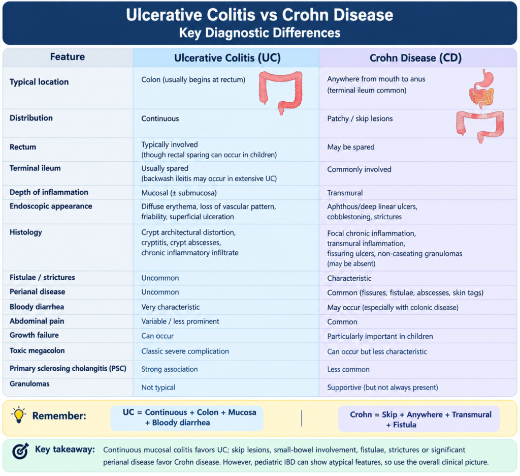
| Feature | Ulcerative Colitis (UC) | Crohn Disease (CD) |
|---|---|---|
| Typical location | Colon (usually begins at rectum) | Anywhere from mouth to anus (terminal ileum common) |
| Distribution | Continuous | Patchy / skip lesions |
| Rectum |
Typically involved (though rectal sparing can occur in children) |
May be spared |
| Terminal ileum |
Usually spared (backwash ileitis may occur in extensive UC) |
Commonly involved |
| Depth of inflammation | Mucosal (+ submucosal) | Transmural |
| Endoscopic appearance | Diffuse erythema, loss of vascular pattern, friability, superficial ulceration | Aphthous/deep linear ulcers, cobblestoning, strictures |
| Histology | Crypt architectural distortion, cryptitis, crypt abscesses, chronic inflammatory infiltrate | Focal chronic inflammation, transmural inflammation, fissuring ulcers, non-caseating granulomas (may be absent) |
| Fistulae / strictures | Uncommon | Characteristic |
| Perianal disease | Uncommon | Common (fissures, fistulae, abscesses, skin tags) |
| Bloody diarrhea | Very characteristic | May occur (especially with colonic disease) |
| Abdominal pain | Variable / less prominent | Common |
| Growth failure | Can occur | Particularly important in children |
| Toxic megacolon | Classic severe complication | Can occur but less characteristic |
| Primary sclerosing cholangitis (PSC) | Strong association | Less common |
| Granulomas | Not typical | Supportive (but not always present) |
Exam Pearls
Pearl 1
UC = continuous colonic mucosal inflammation, typically beginning at the rectum.
Pearl 2
Crohn disease = patchy/transmural inflammation that may involve any part of the gastrointestinal tract.
Pearl 3
Skip lesions, fistulae, strictures and significant perianal disease strongly favor Crohn disease.
Pearl 4
Non-caseating granulomas support Crohn disease but are not required for diagnosis.
Pearl 5
Crypt abscesses are not pathognomonic for UC.
Pearl 6
Growth failure or delayed puberty may be an early manifestation of pediatric Crohn disease.
Pearl 7
Fecal calprotectin is a marker of intestinal inflammation, not a standalone diagnostic test for IBD.
Pearl 8
Rectal sparing does not automatically exclude UC in a child because atypical pediatric UC exists.
Pearl 9
Backwash ileitis can occur with extensive UC and should not automatically be interpreted as Crohn disease.
Pearl 10
PSC is strongly associated with IBD, particularly UC.
One-Minute Board Review
Ulcerative colitis
Rectum → continuous → colon → mucosal → bloody diarrhea → urgency/tenesmus → toxic megacolon → PSC
Crohn disease
Anywhere → skip lesions → terminal ileum common → transmural → abdominal pain → growth failure → perianal disease → fistula/stricture
The safest examination rule
Continuous mucosal colitis favors UC; skip/transmural disease, small-bowel involvement, fistulae, strictures or significant perianal disease favor Crohn disease.
However, do not use a single feature in isolation to classify pediatric IBD because atypical UC, Crohn colitis and IBD-unclassified can overlap.
References
1. Levine A, Koletzko S, Turner D, Escher JC, Cucchiara S, de Ridder L, et al. ESPGHAN revised Porto criteria for the diagnosis of inflammatory bowel disease in children and adolescents. J Pediatr Gastroenterol Nutr. 2014;58(6):795-806. doi:10.1097/MPG.0000000000000239.
2. Wine E, Aloi M, Van Biervliet S, Bronsky J, Martín de Carpi J, Gasparetto M, et al. Management of paediatric ulcerative colitis, part 1: ambulatory care—an updated evidence-based consensus guideline from the European Society of Paediatric Gastroenterology, Hepatology and Nutrition and the European Crohn’s and Colitis Organisation. J Pediatr Gastroenterol Nutr. 2021;73(2):271-291. doi:10.1097/MPG.0000000000003151.
3. Shouval DS, Hojsak I, Miele E, Sokollik C, Turner D, Kolho KL, et al. Management of pediatric Crohn’s disease: an ECCO-ESPGHAN guideline update. J Crohns Colitis. 2026;20(8):jjag084. doi:10.1093/ecco-jcc/jjag084.
4. van Rheenen PF, Aloi M, Assa A, Bronsky J, Escher JC, Fagerberg UL, et al. The medical management of paediatric Crohn’s disease: an ECCO-ESPGHAN guideline update. J Crohns Colitis. 2021;15(2):171-194. doi:10.1013/ecco-jcc/jjaa161.
