Clinical Scenario:
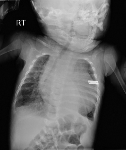
A 3-month-old previously well infant presents with episodes of inconsolable crying, sweating, and pallor during feeding, along with poor weight gain. On examination: HR 160/min, RR 56/min, and a soft apical systolic murmur. ECG shows deep, wide Q waves with ST elevation in leads I and aVL. Chest X-ray shows cardiomegaly. Echocardiography reveals a dilated left ventricle with reduced ejection fraction, moderate mitral regurgitation, and retrograde flow noted in a dilated right coronary artery.
What is the most likely diagnosis?
A. Anomalous left coronary artery from pulmonary artery
B. Dilated cardiomyopathy
C. Kawasaki disease with coronary aneurysm
D. Myocarditis
E. Supravalvular aortic stenosis
Correct answer & Explanation:
Correct answer: A. Anomalous left coronary artery from the pulmonary artery (ALCAPA)
This is a classic presentation of ALCAPA (Bland–White–Garland syndrome) in an infant. The combination of age 2–4 months, feeding-related distress/diaphoresis, poor weight gain, cardiomegaly, LV systolic dysfunction, mitral regurgitation, and ischemic ECG changes is highly characteristic.
Why ALCAPA is the most likely diagnosis
In ALCAPA, the left coronary artery arises abnormally from the pulmonary artery instead of the aorta.
Immediately after birth, pulmonary arterial pressure is relatively high, so coronary perfusion may initially be adequate. Over the following weeks, pulmonary vascular resistance and pulmonary artery pressure fall. This creates a coronary steal phenomenon: blood can flow retrogradely from the coronary circulation toward the low-pressure pulmonary artery rather than adequately perfusing the left ventricular myocardium. This produces myocardial ischemia and infarction.
This explains the characteristic age of presentation. Infants commonly become symptomatic around 2–4 months, when pulmonary vascular resistance has fallen sufficiently.
Clinical clues in this question
1. Age — 3 months
This is a classic age for symptomatic infantile ALCAPA. The infant is initially well and then develops symptoms as pulmonary vascular resistance falls.
2. Episodes of inconsolable crying, pallor and sweating during feeding
These episodes represent infantile angina caused by myocardial ischemia. Feeding increases myocardial oxygen demand, making ischemic symptoms more apparent.
ALCAPA may therefore be mistaken for colic, reflux or feeding intolerance in infancy.
3. Poor weight gain
Chronic myocardial ischemia and heart failure result in feeding difficulty and failure to thrive.
4. Deep, wide Q waves with ST elevation in I and aVL
This is a particularly important clue.
ALCAPA characteristically produces anterolateral myocardial ischemic/infarction changes, including prominent/deep Q waves in lead I, aVL and left precordial leads.
Therefore, an infant with heart failure plus deep Q waves in I and aVL should immediately raise suspicion for ALCAPA.
5. Dilated left ventricle with reduced ejection fraction
Chronic myocardial ischemia causes LV myocardial dysfunction and dilatation. Infantile ALCAPA commonly presents with significant LV systolic dysfunction.
6. Mitral regurgitation
This is another important clue. Ischemia can affect the papillary muscles and mitral valve apparatus, producing mitral regurgitation. LV dilatation can further worsen functional MR.
7. Dilated right coronary artery with retrograde coronary flow
This is perhaps the most diagnostic echocardiographic clue.
Because the left coronary system is poorly perfused, collateral vessels develop from the right coronary artery (RCA). The RCA may become enlarged and supply collateral blood toward the left coronary circulation. Ultimately, blood can pass through the anomalous coronary circulation and drain into the pulmonary artery, producing the characteristic coronary steal.
Therefore, a dilated RCA with abnormal/retrograde coronary flow in an infant with LV dysfunction is highly suggestive of ALCAPA.
Why the other options are wrong
B. Dilated cardiomyopathy — Incorrect
Dilated cardiomyopathy can certainly produce:
- Cardiomegaly
- LV dilatation
- Reduced ejection fraction
- Heart failure
- Mitral regurgitation
So it is an important differential diagnosis.
However, it does not explain the deep ischemic Q waves with ST elevation and the abnormal coronary anatomy/flow.
Most importantly, the presence of a dilated right coronary artery with abnormal coronary flow points toward a coronary anomaly rather than primary myocardial disease.
In an infant with apparent dilated cardiomyopathy, ALCAPA should specifically be excluded, because it is a potentially correctable cause of LV dysfunction.
(If you like , solve the MCQ on dilated cardiomyopathy)
C. Kawasaki disease with coronary aneurysm — Incorrect
Kawasaki disease can produce coronary artery aneurysms, but this diagnosis is extremely unlikely in this clinical setting.
The infant has a congenital coronary anomaly with infantile ischemic heart failure, rather than the typical clinical picture of Kawasaki disease.
Kawasaki disease would generally have a history of prolonged fever and systemic inflammatory features. None are present here.
Furthermore, the combination of deep Q waves, LV dysfunction and ischemic MR in a 3-month-old is much more characteristic of ALCAPA.
D. Myocarditis — Incorrect
Myocarditis can cause:
- Tachycardia
- Poor feeding
- Tachypnea
- Cardiomegaly
- LV systolic dysfunction
- Mitral regurgitation
Therefore, it can resemble ALCAPA clinically.
However, myocarditis does not explain the highly characteristic coronary artery abnormality, particularly the dilated RCA and abnormal coronary flow.
The ECG ischemic pattern and echocardiographic coronary findings make ALCAPA much more likely.
E. Supravalvular aortic stenosis — Incorrect
Supravalvular aortic stenosis produces left ventricular outflow obstruction and may be associated with abnormalities of the coronary arteries, particularly in Williams syndrome.
However, this condition would be expected to produce evidence of aortic outflow obstruction, such as a characteristic systolic ejection murmur and an elevated aortic pressure gradient.
It does not explain the classic combination of:
infantile angina + deep Q waves + LV dysfunction + MR + abnormal coronary flow.
Therefore, it is not the best diagnosis.
High-yield postgraduate exam clue
A very useful way to recognize infantile ALCAPA is:
3-month-old infant + sweating/crying during feeds + poor growth + cardiomegaly + LV dysfunction + deep Q waves in I/aVL ± mitral regurgitation = think ALCAPA.
The deep Q waves in I and aVL are especially valuable because they indicate anterolateral myocardial infarction/ischemia, which is a classic ECG clue for ALCAPA.